
Cell Gallery
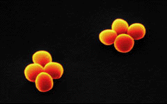 |
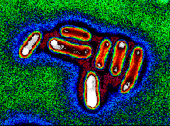
A pair of Staphylococcus aureus have gone through 2 cell divisions, producing a pair of tetrads. Cell division in this and other bacteria can occur every 20 to 30 minutes. Elsewhere on this site, you can learn about the dynamics of bacterial growth. |
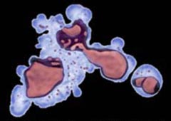 |
This leukemia cell is tearing itself apart by a process called apoptosis. You can find out more about this natural phenomenon of programmed cell death in "Apoptosis: when a cell commits suicide". |
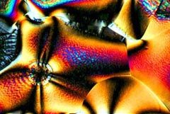 |
Ascorbic acid (vitamin C) was dissolved in ethanol and allowed to recrystallize through evaporation. The crystal was photographed between crossed polarizing filters. Other crystals are on exhibit in "CRYSTALS alive!" |
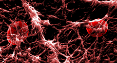 |
This scanning electron micrograph shows the fine structure of a blood clot. Platelets released from the circulation and exposed to the air use fibrinogen from the blood plasma to spin a mesh of fibrin. Learn how other cells defend us against infection in "OUCH!... Anatomy of a Splinter". |
 |
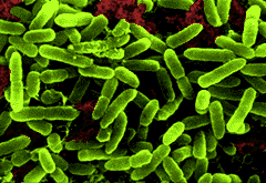
Escherichia coli (E. coli ) are very common intestinal inhabitants. Some can be dangerous in food and water supplies. Most newsworthy are life-threatening infections from eating undercooked E. coli 0157 - contaminated hamburger. Before leaving "CELLS alive!", learn how bacteria swim and how they grow. |
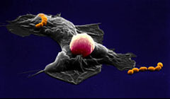 |
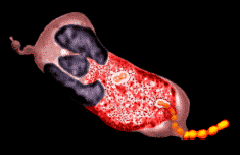
Scanning electron micrograph of human macrophage ingestingStreptococcus pyogenes. The spherical cell riding piggy-back on the macrophage is a lymphocyte, an important component in the immune response to infection. Read about these cells' close association in Antibody Production. |
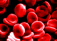 |
Scanning electron micrograph of human red blood cells. Red cells get their red color from iron-rich hemoglobin which is responsible for transporting oxygen throughout the body. These cells got THEIR red color from Photoshop(R). See animated red cells in capillaries in "OUCH!... Anatomy of a Splinter". |
 |
Scanning electron micrograph of the common soil bacteriaPseudomonas aeruginosa. These bacteria are actively motile in aqueous environments but can attach to a submerged surface and grow into a sessile, slimy colony called a "biofilm". Colorized gray scale image. |
 |
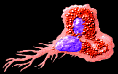
Human neutrophils are white blood cells that serve as professional phagocytes: their primary function is to eat and kill bacteria and they arrive quickly at the site of a bacterial infection. This neutrophil, ingesting Streptococcus pyogenes, was imaged in gray scale with phase contrast optics and colorized. See neutrophils in action in "OUCH!... Anatomy of a Splinter". |
 |
This human eosinophil was originally imaged in gray scale with Nomarski DIC optics and a CCD camera, then colorized using Adobe Photoshop(R). Eosinophils are important in combatting parasitic diseases and people with those diseases and allergies generally have elevated eosinophil counts. See Allergies and Mites. |